
Complicated orthodontic cases require more information for the orthodontist to derive optimal diagnosis than simple cases do (Mah et al., 2010).
Figure 8.5 illustrates a complicated orthodontic case consisting of two fused maxillary incisors and some supernumerary teeth. Due to the amount of crowding, it is difficult to ascertain the locations of the supernumerary teeth and the quality of their roots. A CBCT scan taken to determine the best treatment plan for the patient revealed that a supernumerary right maxillary lateral incisor was located palatal to the fused teeth and a supernumerary left maxillary central incisor was located distal and apical to the erupted left maxillary central incisor (Figure 8.6A,B).
To give further context to the positions of the supernumerary teeth relative to the soft tissue, Adobe Photoshop® was used to superimpose the patient’s maxillary occlusal intraoral photograph onto a screenshot of the maxillary dentition rendered from the CBCT scan (Figure 8.6C) and the information used to derive an well-informed treatment plan (Figure 8.6D).

Figure: 8.5 A: Panoramic radiograph, C : Maxillary occlusal intraoral
Figure 8.5 A: Panoramic radiograph showing two fused maxillary right incisors and several supernumerary teeth. B: Frontal intraoral photograph showing fused teeth. C: Maxillary occlusal intraoral photo showing fused teeth and erupting supernumerary teeth.
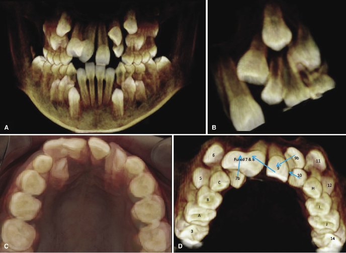
Figure 8.6 A: Frontal view of volume rendering showing fused teeth.
The comprehensive treatment plan depicted in Figure 8.6D involved the extraction of the fused teeth (labeled Fused 7 & 8) followed by the bonding of fixed edgewise-orthodontic appliances. The supernumerary right lateral incisor (labeled 7b) then would be brought labially and the erupted left central incisor (labeled 9) moved mesially across the midline to replace the extracted fused teeth. The supernumerary left central incisor (labeled 9b) then would be moved inferiorly and mesially to replace the left central incisor that had been moved across the midline. At a future date, the erupted left central incisor would require an esthetic bonding or veneer to give it the appearance of the missing right central incisor. This treatment plan would allow the patient to keep a full complement of teeth without the need for dental implants or a fixed prosthesis. After 18 months of active orthodontic treatment, the panoramic radiograph shows the improved positions of the teeth (Figure 8.7A); after 22 months of treatment, the dramatic changes could be appreciated clinically (Figure 8.7B,C).

Figure 8.7 A: Panoramic radiograph
Figure 8.7 A: Panoramic radiograph showing treatment progress at 18 months. B and C: Maxillary occlusal and frontal intraoral intraoral photograph, respectively, showing treatment progress at 22 months with the orthodontist taking photos to show the progress made.
Fusion Radiology provides Dental CBCT scan reporting service by NHS Dental radiologists, for further information please email info@fusion-radiology.com, call 01582 249449, 07828634357


