
Collecting diagnostic information: CBCT
The review of a CBCT scan is a step-by-step analysis of all abnormal radiographic findings or features with the goal of recognizing and collecting as much information as possible that is available in the various image reconstructions (White & Pharoah, 2004). Analysis of the collected information may lead the clinician to a diagnosis or, more frequently, to a short list of possible diagnoses.
As a first step, the disease or the abnormality has to be identified. For this purpose, the diagnostician should look for irregularities in the appearance of the region that the clinical examination and/or other findings have indicated as the area of concern, including changes in the shape and size or density affecting hard or soft tissue or both at the site. Alterations in the cortical or cancellous bone pattern (Figure 9.1 and Figure 9.2) are a frequent result of disease. Although dentists largely are familiar with normal and abnormal appearance of the osseous tissue in traditional dental diagnostic images, getting acquainted with irregular appearance of tissues in CBCT may require substantial exposure to and experience with these images (Ahmed et al., 2012). With regard to soft tissues, CBCT images show minimal differences in density of various soft tissues (Figure 9.3); their changes in disease processes are almost impossible to identify unless the disease has caused either extensive destruction of the soft tissue involved or has induced the production of dystrophic calcifications.
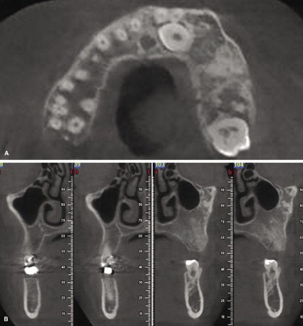
Axial (A) and cross-sectional (B) images, maxillary bone
Figure 9.1 Axial (A) and cross-sectional (B) images of the maxilla depicting an asymmetry between the right and left side of the maxillary bone. Note the difference in the density of the alveolar bone between the left and right sides. This is the kind of information to be documented when reviewing the region of interest.

Axial (A), cross-sectional (B), and panoramic (C) reconstruc
Figure 9.2 Axial (A), cross-sectional (B), and panoramic (C) reconstruc.
Fusion Radiology provides Dental CBCT scan reporting service by NHS Oral and Maxillofacial Radiologist, for further information please email info@fusion-radiology.com, call 01582 249449, 07763875191


